Tuesday 11 June 2013

Ovarian Tumours

Ovarian tumours in dogs and cats are very uncommon. True reports of these tumours are unknown because most reports in literature are based on necropsy findings. The reason behind low true clinical evidence of ovarian tumours is large segment of canine and feline population is surgically neutered at an early age.
                                                                      Ovarian tumours are mainly classified into three categories, according to nature of cell origin.
·        


  Epithelial cell


·          Germ cell
·          Sex cord stromal cell
Breeds which get affected in common are Pointers, English Bulldogs, Boxers, German Shepherds, Yorkshire Terriers and Indian Hounds.
1)        Epithelial Cell Tumours-
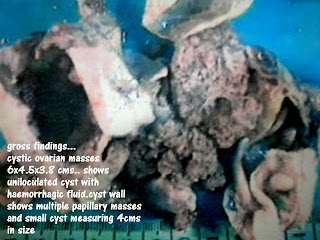
These tumours mainly include papillary adenoma, papillary adenocarcinoma, papillary cyst adenoma and adenocarcinoma. About 40 to 60% of canine ovarian tumours fall in this category. Papillary adenocarcinoma often is associated with widespread peritoneal invasion and marked by malignant haemorrhagic effusion often misleading to ascites.
Development of malignant effusion is due to
i) Leakage of fluid through tumour capsule.
ii) Exfoliation of tumour cells resulting in transcoelomic metastatic implants that exert pressure and obstruct peritoneal and diaphragmatic lymphatics.
iii) Secretion from metastatic peritoneal implants
Papillary adenocarcinoma usually metastasizes to kidney, liver, lungs, and omentum and par aortic lymph nodes.
Cystadenocarcinomas originate from the rete ovarii and consists of multiple thin walled cysts.
2)        Germ Cell Tumours-
The ovarian primordial germ cells are responsible for ovarian        dysgerminomas, teratomas and teratocarcinomas.
Dysgerminomas arise mainly from undifferentiated germ cells and consists of ovarian primordial cells. Due to their resemblance with testicular cells they are also called as “ovarian seminomas”. These tumours grow by expansion and metastasis is often in abdominal lymph nodes. However involvement of other vital organs has been also seen.
Teratomas are mainly composed of germ cells which are differentiated in two or more germinal cell layers. So these tumours have both mature elements and undifferentiated elements resembling those of embryo. These are highly metastatic in nature and it occurs in all other vital body parts.
3)        Sex Cord Stromal Tumours-
These tumours arise from the specialised gonadal stroma of the ovary, which is responsible for oestrogen and progesterone production. Most common sex cord stromal tumour is “Granulosa Cell Tumour”.
These are firm, lobulated and grow quite large size, have good metastatic potential. They are quite potential to elaborate steroid hormones.
Alpha-inhibitin, a gonadal glycopeptides known to be feedback inhibitor of pituitary secretion of follicular stimulating hormone (FSH), is a useful histologic marker of Granulosa cell tumour.
The other stromal cell tumours like Thecomas and Luteomas are also reported and are benign in nature.
Other tumours like conditions which are found are par ovarian tumours which originate from mesonephric tubules, cystic rete tubules, vascular haematomas and adenomatous hyperplasia of rete ovarii.
Depending upon the tissue of origin the clinical symptoms of ovarian tumour vary. Main clinical symptom of epithelial cell tumours is malignant ascites. Germ cell tumours are associated with hormonal dysfunction and space occupying mass in abdomen. Routine abdominal radiographs show enlarged mass with calcified foci. Stromal tumours secrete steroid hormones hence excessive oestrogen hormone leads to vulvar enlargement, sanguineous vulvar discharge, persistent oestrous, alopecia and aplastic pancytopenia. Excessive progesterone production leads to cystic endometrial hyperplasia/ pyometra complex.
The common diagnostic techniques are abdominal radiography and ultrasonography. Cytological evaluation of the malignant effusion from abdomen gives better idea.
Palliative radiation and chemotherapy using platinum based combinations with taxanes is useful in human patients but radical surgery remains the mainstay of treatment for ovarian tumours in canines.
Complete ovariohysterectomy is recommended. During surgery you have to gentle in handling tissues to minimise transcoelomic tumour spread. Before abdominal closure careful examination of all serosal surfaces, including omentum, diaphragm is necessary. Any suspected lesions should be subjected to biopsy, FNAC for metastatic disease. Before closure of abdominal incision a through wash with ample of NS is advisable.
Prognosis is very good after surgery if no metastasis and adhesions are noticed.


Wednesday 10 April 2013


FIBROSARCOMA


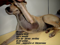
Fibro sarcomas are classified as soft tissue sarcomas which are heterogeneous group of tumours. They mainly arise from mesenchymal tissues and have features similar to the cell type of origin.

These tumours originate in connective tissues like muscle, adipose, neurovascular, facial, fibrous tissue and are benign or malignant entities. They may arise from any anatomical site in body but skin and subcutaneous sites are most common.

All these fibro sarcomas have following features

1. They appear as pseudo encapsulated soft to firm tumours of poorly defined histologic margins

2. They are locally invasive and infiltrate through and along facial planes

3. After conservative surgical excision recurrence is common.

4. Haematogenous metastasis is up to 20% of cases but regional lymph node metastasis is unusual.

5. Metastasis depends on histopathologic grade of the tumour and resected tumour margins predict local recurrence.

6. Tumours more than(>5) cm in diameter have poor response to chemotherapy and radiation therapy.

Development of immunocytochemical procedures, the availability of monoclonal and polyclonal antibodies to various tissue markers, tissue micro array technology improved diagnosis of soft tissue sarcomas is human pathology but it is still limited degree in veterinary pathology. Usually all these tumours are graded as I, II, III, IV with ascending intensity.

Most of the fibro sarcomas arise from skin and subcutaneous tissue and represented with malignant fibroblasts. They are well differentiated and exhibit spindle shaped tumour cells with scanty cytoplasm. More anaplastic tumours show dense fibroblasts with mitosis and pleomorphism.

All fibro sarcomas tend to occur in old dogs and cats without sexual predilection.Retrieviers and Dobermans are more prone for fibro sarcomas.

All these tumours are easily diagnosed with FNAC, biopsy techniques. Routine haematological and serum biochemistry do not show any changes except anaemia and thrombocytopenia in few cases.

Modern three dimensional imaging techniques such as digital radiography, CT, MRI and ultrasonography are very useful diagnostic tools available now days and they are very useful to decide surgical approach and radiation therapy protocol.

Modified AJCC staging system is used for histological grading and clinical staging (TNM system) of fibro sarcoma.

Most of the fibro sarcomas are treated either by surgical resection, radiation and chemotherapy or suitable combination of them. Palliative radiation up to 30 to 45 GY. Is sufficient. Doxorubicin based protocols are considered as best for chemotherapy.


Friday 29 March 2013

CANINE  LYMPHOMA

              The lymphoma (malignant lymphoma or lymphosarcoma ) are a diverse group of neoplasms which originates from lymphoreticular cells. 
             They usually arise in lymphoid tissues like lymph nodes, spleen and bone marrow. However they may arise in almost any tissue in the body. Lymphoma one of the most common neoplasms in dogs and is one of major (5th cause ) 'cause of canine mortality. Non-Hodgkin's lymohoma accounts 5% of all new cancer cases in canines.
                Dog breeds which are susceptible to lymphoma are Boxers, Retrievers, Bull Mastiffs, Basset Hounds, Saint Bernards, Scottish terriers, Airedales, Bulldogs. Breeds which are less susceptible are Dash-hounds and Pomeranians.
                Etiology -is unknown and multifactorial. Broadly factors are 1). Genetic and molecular
2). Infectious 3). Environmental ( herbicides and  strong magnetic fields) 4). Immunosuppression 
                Lymphomas are classified by various systems out of which (NHC), HCI updated  Kiel system and revised EAL systems are important. But WHO' s clinical staging system for lymphosarcoma in domestic animals is the most followed system. 
                Hypercalcemia and anemia, with CNS depresion are considered to be pathognomonic symptoms. Complete Blood count, biochemistry, urine analysis, Histological and cytological evaluation of lymph nodes  with FNAC technique, Molecular diagnostic techniques (PCR), and clonality assay are methods used for clinical staging  of diasease .
               Lymphoma are treated with radicle surgery followed by palliative radiation, multidrug or single drug chemo therapy for which lymphoma protocol is available. 
               Although canine lymphoma has rarely curable, prognosis mainly varies and depends on number of factors such as location of disease, extent of disease, presence or absence of clinical signs, the histologic grade, the immunophynotype  (T cell or B cell ), exposure to previous chemotherapy or corticosteroids and subsequent development of MDR, altered cell death processes ( apoptosis), the proliferation rate of the tumor, the presence of paraneoplasitc conditions and possible gender.      


LARYNGEAL TUMORS

Laryngeal Tumors are rare in nature and include Rhabdomyoma (oncocytoma ), osteosarcoma, extramedullary plasma cytoma, chondrosarcoma, undifferentiated carcinoma fibrosarcoma, mastcell tumors, adenocarcinoma and sqamous cell carcinoma.
         Patients with laryngeal carcinoma usually have a progressive change in voice or bark,exercise intolerance or dysphagia.
        Diagnosis of such tumors is very easy because of easy direct biopsy, FNAC and digital radiography.
        Benign laryngeal tumor of cysts can be removed surgically with better results. But in malignancy laryngectomy with permanent tracheostomy which is best options for humans can not be undertaken in canines. 
         Radioresponsive tumors can be irradiated with palliative rediotherapy with 35 to 45 Gy. radiation potential. Benign tumors have good prognosis. Unfortunately very limited information is available for malignant tumors and their treatment because very few have been treated and reported. 
        Doxirubicin is a drug of choice for chemotherapy and palliative  radiation with 32 to 45 Gy. can pull patient's life upto 1 year maximum.


Sunday 17 March 2013

Peripheral Nerve Sheath Tumors.

(PNST)


PNST are malignant tumors of nerve sheath origin and have been referred to as neurofibrosarcoma, malignant schwannoma and haimangiopericytoma. They occur any where in body. PNST are divided mainly in three groups 
1. Peripheral group -  PNST involves nerves away from brain or spinal cord.
2. Root group - Involves nerves immediately adjescent to brain or spinal cord
3. Plexus group - involves brachial or lumbosacral plexsus
       True peripheral forms of PNST are easily treatable.
PNST are poorly defined tumors and appear like fibrosarcomas. They are adherent to deeper tissues and infiltrate underlying fascia,muscle and skin.
        All PNST's are malignant but have moderate metastetic rate. Recurrence after conservative surgery is very common. modified staging and grading system for these PNST is available.
         The best chemotherapeutic agent is doxorubicin and palliative radiation at the rate of 30 Gy usually undertaken.Survival is 12 to 24 months maximum, reported after diagnosis.